From nanoparticle to biomarker...
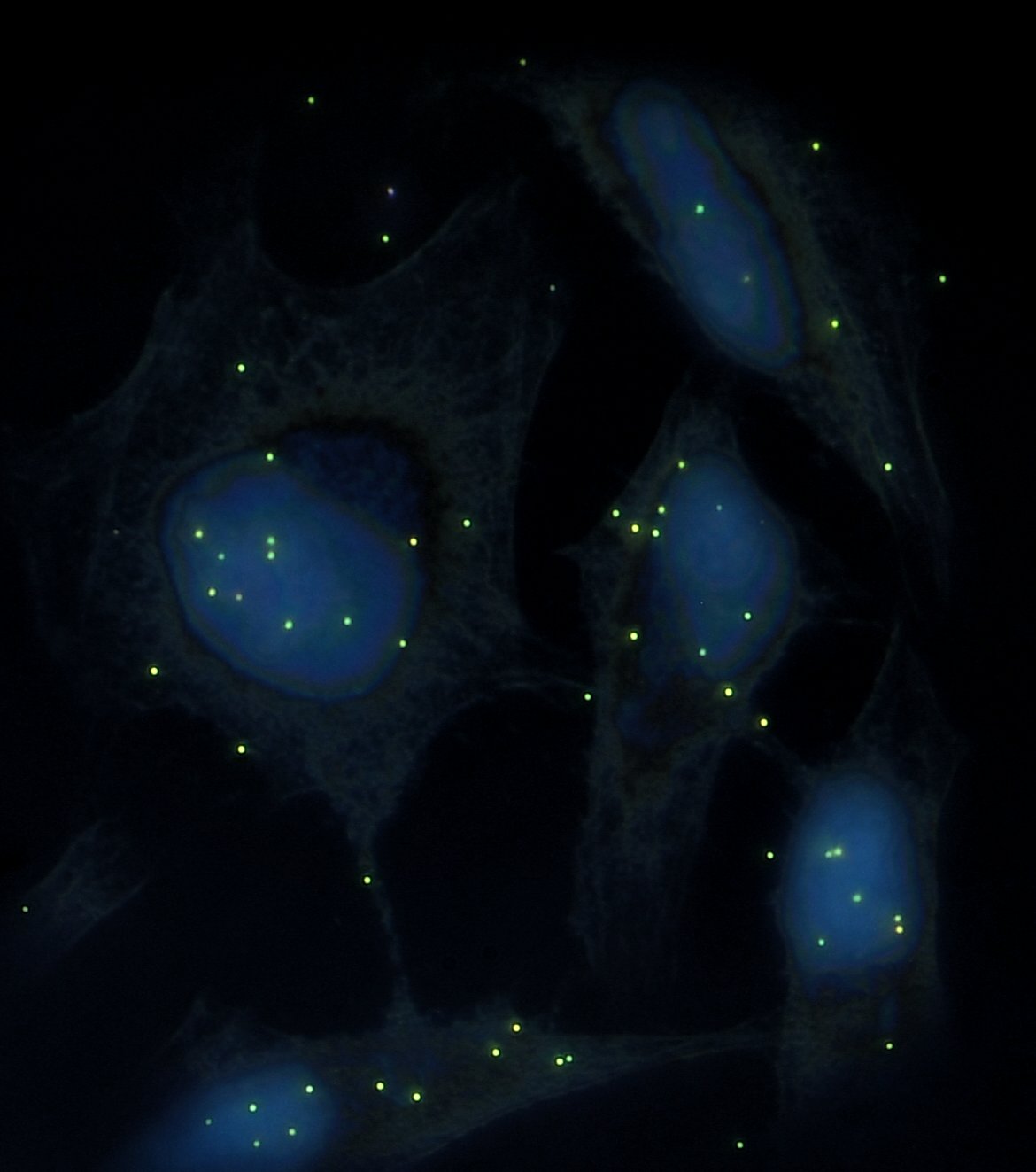
The surface modification of the nanoparticles by attaching specific antibodies transforms the nanoparticles into biomarkers, which will target the cells carrying the antigens that we want to identify. The use of nanoparticles of different colors, functionalized with different antigens, thus opens the way to quantitative multiplexing in cytopathology. We propose to use here four markers, ranging from blue to red, composed of gold-silver alloys or gold only. Counting the number of markers attached to the cell membranes should thus make it possible to quantify the level of expression of the antigens on the cell surface and thus determine whether they are healthy or malignant cells.
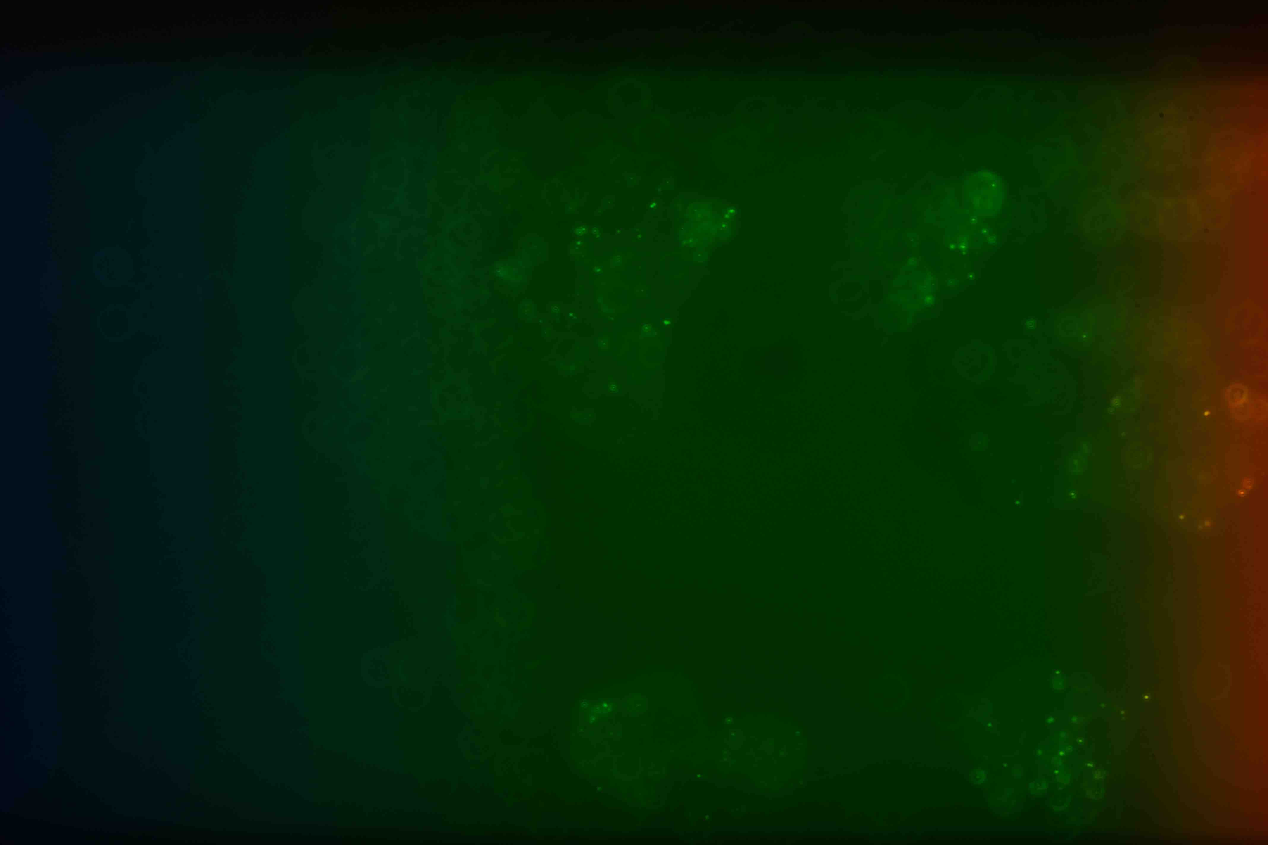
HYPERSPECTRAL imaging in PATHOLOGy
Hyperspectral imaging consists of the analysis of the different wavelengths diffused or transmitted by a sample. It allows us here to identify the different plasmonic nanoparticles used with the help of their light scattering spectrum. This is to propose a method of rapid acquisition and accurate classification of malignant vs benign cells within cytopathological sample.
Machine learning will save the world!

Machine learning is proving to be a great tool for classifying and segmenting images. It is with a neural network (deep learning) that we want to achieve the segmentation of the images obtained with the hyperspectral camera and to classify and quantify the number of nanoparticles per cell. The goal is to identify with great reliability the malignant cells and healthy cells from a patient.
BIBLIOGRAPHY
- Rioux D, Meunier M, Seeded growth synthesis of composition and size-controlled gold-silver alloy nanoparticles, Journal of Physical Chemistry C, 2015.
- Bergeron É, Patskovsky S, Rioux D, Meunier M, 3D multiplexed immunoplasmonics microscopy, Nanoscale, 2016.
- Qi M, Darviot C, Patskovsky S and Meunier M, Cost-effective side-illumination darkfield nanoplasmonic marker microscopy, The Analyst, 2019.